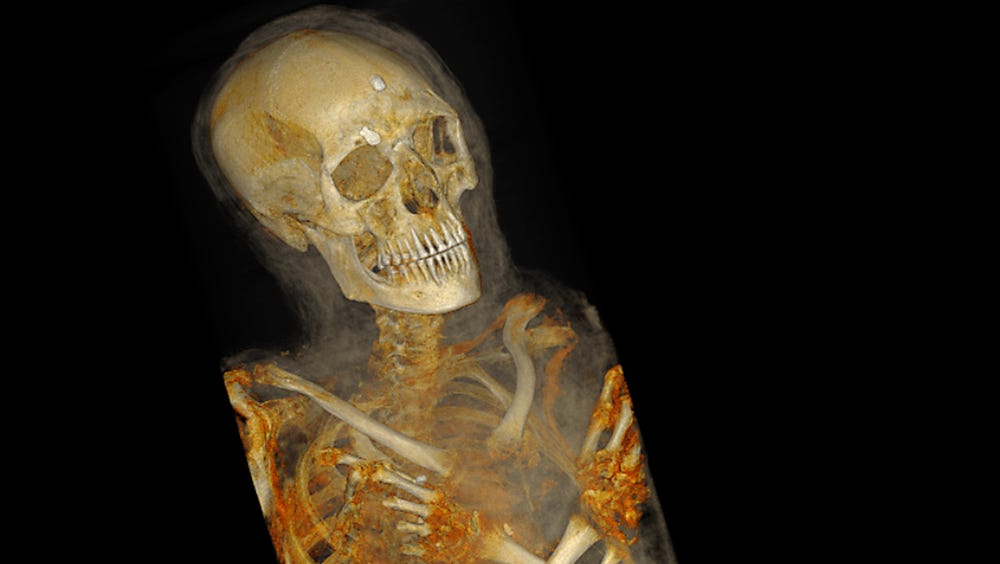
Very Postmortem: Mummies and Medicine Opens at the Legion of Honor on Halloween 2009
Oct 1, 2009
SAN FRANCISCO—Archaeology meets technology in Very Postmortem: Mummies and Medicine which opens on Halloween (October 31, 2009) in Gallery 1 at the Legion of Honor Museum in San Francisco. This exhibition welcomes back to the Fine Arts Museums the mummy of Irethorrou, a priest from an important family who lived in Akhmim, Egypt around 500 B.C., died at a young age and was buried over 2,500 years ago. The mummy had been on long-term loan to the Haggin Museum in Stockton, California for the last 65 years. Using state of the art High Definition Volume Rendering® technology by Fovia, Inc., the exhibition reveals Irethorrou’s long held secrets through three-dimensional computed tomography (CT) scans conducted by the Stanford Medical School’s Department of Radiology and presents a forensic portrait reconstructed by the Akhmim Mummy Studies Consortium.
“What we’re trying to do is merge science, culture, history, medicine and art. This exhibition gives us an opportunity to incorporate modern techniques and procedures with one of the oldest objects in our permanent collection,” describes Dr. Renee Dreyfus, curator of ancient art and interpretation for the Fine Arts Museums of San Francisco.
To the ancient Egyptians, the preservation of the body was an important factor in attaining and maintaining an afterlife. Mummification evolved from the concept of preserving the body as a receptacle for the life force, which survives after death. Although great numbers of mummies were exported as “curiosities,” they have been an underestimated and underutilized resource that is finally becoming recognized as a rich trove of preserved material. Many mummies from the Egyptian city of Akhmim were found and exported at the end of the 19th century allowing scientists to study them as a population. Today modern scientific examinations of these relics are providing exciting new insights into the conditions under which these individuals lived, bringing us even closer to an understanding of who they were.
Very Postmortem: Mummies and Medicine examines the ancient practice of mummification through the lens of modern technology. While the mummy of Irethorrou lies in its decorated coffin, visitors can learn how modern mummy research is advanced through the use of high-resolution scanners. Images produced by the Akhmim Mummy Studies Consortium and by scientists at Stanford Medical School’s Department of Radiology and Fovia, Inc. reveal much about Irethorrou and how he was prepared for eternity, including the locations and textures of over a dozen magic amulets that were placed on his body during the intricate wrapping process.
The exhibition will include computer-generated models of the skulls of Irethorrou and of a close relative Ankh-Wennefer, owned by the Washington State History Museum in Tacoma, Washington. This affords the rare opportunity to reunite members of the same ancient family through forensic portraiture.
“Through this exhibition, we hope to create an historical reconstruction of Irethorrou’s life as an ancient Egyptian,” explains Dr. Jonathan Elias, director of the Akhmim Mummy Studies Consortium.
Very Postmortem: Mummies and Medicine includes other “cult of the dead” antiquities that relate to the ancient Egyptian beliefs of death and the afterlife including a beaded mummy mask from Dynasty 26 (7th century B.C.), an anthropoid coffin from Dynasty 30 (4th century B.C.), a funerary shroud circa A.D. 180-275, amulets, funerary furnishings and a selection of historical prints that highlight the public’s ongoing fascination with mummies. The exhibition opens on October 31, 2009 and runs through October 31, 2010.
Irethorrou is one of four human mummies and one crocodile mummy in the Fine Arts Museums’ permanent collection. These and other antiquities were among the museum’s earliest gifts, having been given to the collection by founders M.H. de Young, Adolph Spreckels and other donors.
Organization
This exhibition is organized by the Fine Arts Museums of San Francisco with the cooperation of the Akhmim Mummy Studies Consortium and the Radiology Department of the Stanford Medical School. Additional project assistance has been provided by the Stanford Division of Anatomy, ehuman Inc., and Fovia Inc.
Generous support is provided by the William E. Winn, Jr, Living Trust and the Dorothy Tyler Living Trust.
Thank you to Intel Corporation for its generous in-kind donation.
